Türkçe
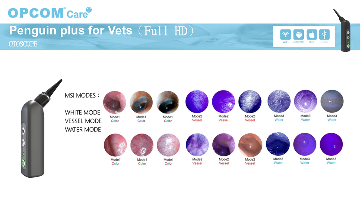
VETERINARY WI-FI (WIRELESS) VIDEO OTOSCOPIC DEVICE
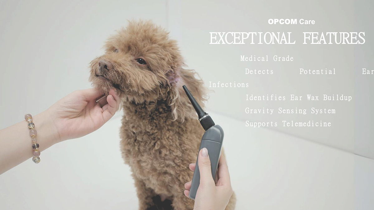
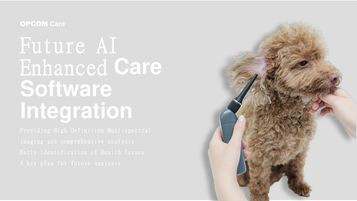
Wi -Fi enabled wireless video otoscope devices for veterinary clinics and field veterinary practice ; for diagnosing inner ear diseases ( otitis) Accurate diagnosis of parasites ( externa , foxtail grass, etc.) and proper communication with pet owners (pre-treatment/post-treatment demonstrations) has become a standard requirement today.
General Operating Principle
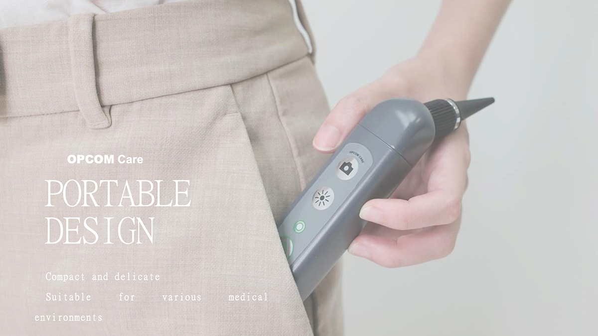
Video otoscope devices transmit real-time HD images and videos wirelessly to tablets, phones ( iOS / Android ), or computers via their built-in Wi -Fi network.
1. FEATURED MODELS AND THEIR SPECIFICATIONS
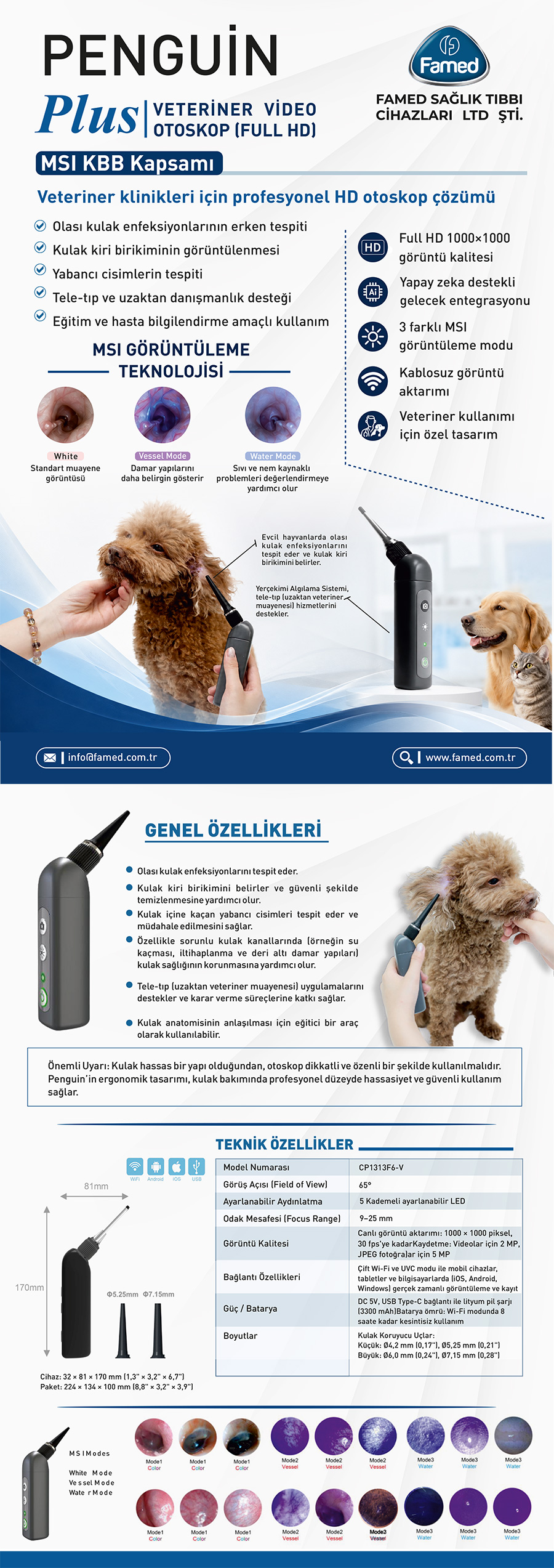
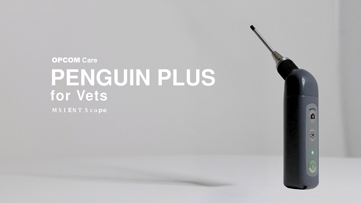
A. OPCOM Penguin Plus ppv (W- LAN HD Digital Otoscope (VET)
Globally and in clinics in Turkey, the veterinary video otoscope is one of the most commonly used professional devices.
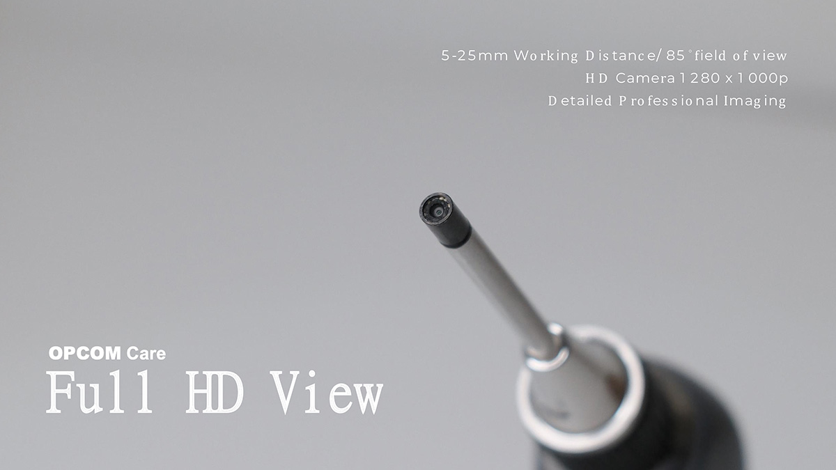
- Camera and Resolution: 1000x1000 pixel HD camera system, 2 MP sensor and 5 MP photo quality.
- Video Recording: Features smooth 30 FPS video recording.
- Viewing and Focusing: Wide 65-degree field of view and 9-25 mm focusing distance.
- Lighting: Fully adjustable, 5-level ultra-bright LED lighting system on the body.
- Connectivity: Fully compatible with mobile devices, tablets, and computers ( iOS , Android , Windows) thanks to dual Wi -Fi bands.
- Battery Life: With its 3300 mAh high-capacity battery, it provides up to 8 hours of uninterrupted operation .
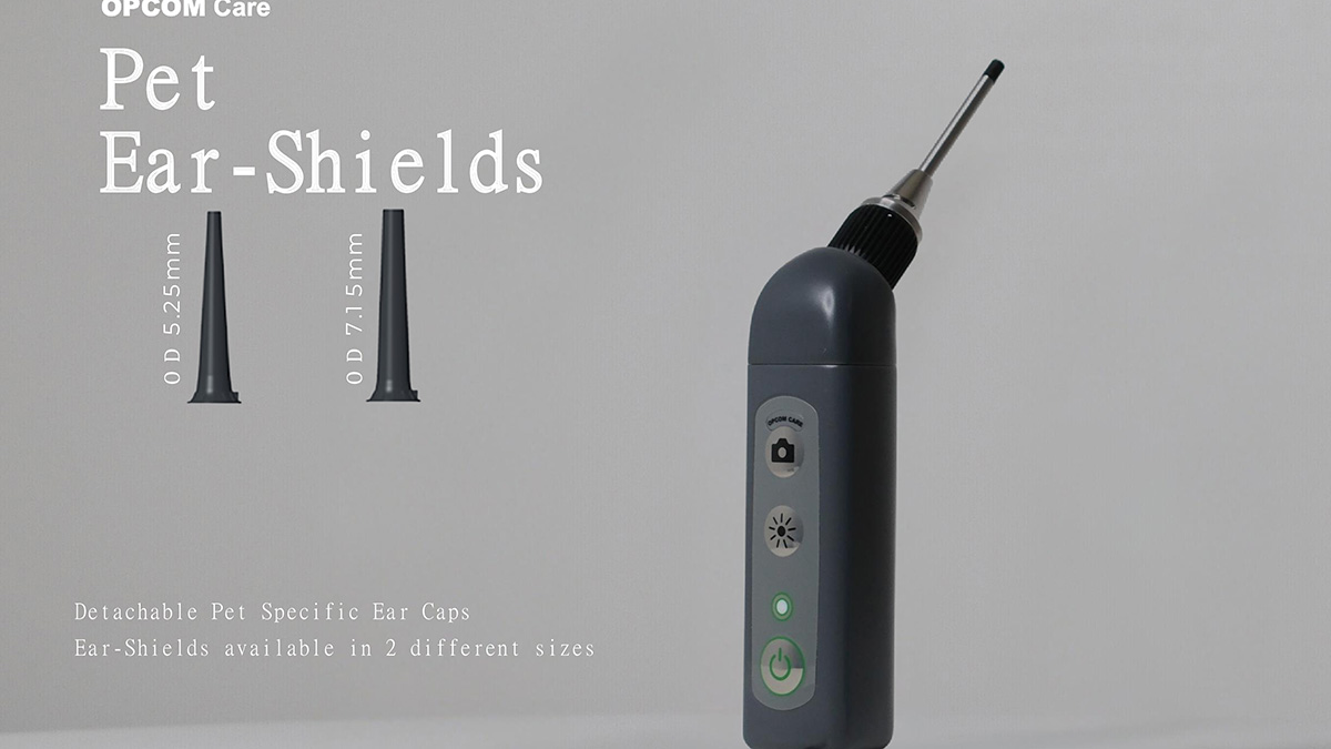
- Ease of Use: The camera body features a shutter button for taking instant photos.
B. Famed Wireless Video Otoscope Device
- frequently preferred by veterinarians in the Turkish market, with a warranty from both domestic and imported distributors .
- It works seamlessly with clinical automation software and mobile devices.
- Earwax analysis is actively used in foreign body diagnosis and patient monitoring.
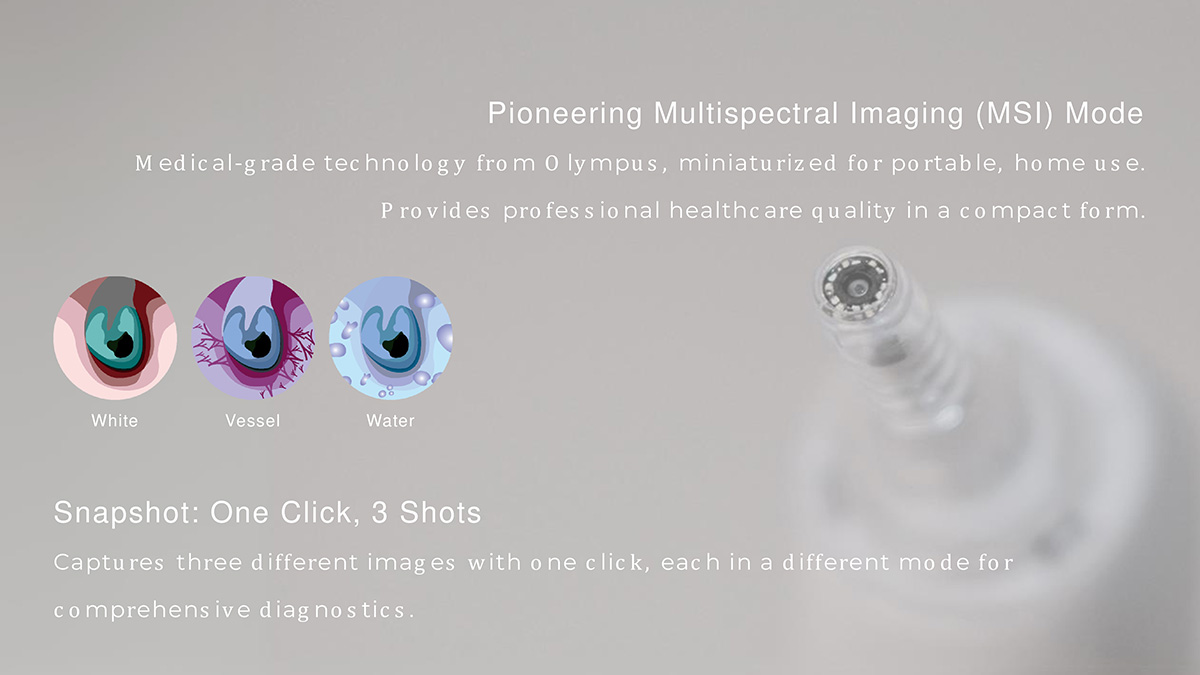
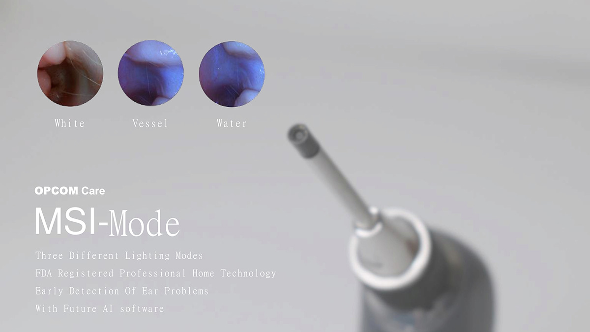
- "THREE SEPARATE LIGHT PROCESSING" (MULTISPECTRAL) TECHNOLOGY
advanced veterinary video otoscopes , endoscopes, and microscopes, utilizes three different light modes to better examine subcutaneous tissue, improve image quality, and differentiate between various pathologies :
| Light Mode | Function / Technical Specification | Intended for Clinical Use |
|---|---|---|
| 1. White Light | It is a standard examination light. It reflects the natural colors and anatomical structure of the tissue in the most realistic way. | Routine checks, general examinations, and detection of obvious foreign bodies. |
| 2. Green Light (NBI) | Narrowband Imaging Band (Imaging ) presents this. The hemoglobin protein found in blood strongly absorbs green light. | the capillary network ( vascularization ), microhemorrhages in the mucosa, tissue damage, tumor formation, and foci of chronic inflammation . |
| 3. Blue / UV Light | It is a special type of short-wavelength light that creates a fluorescent effect and enhances tissue contrast . | With the help of fluorescent agents, it makes bacterial infections , fungal growths, scratches, and ulcers on the cornea or mucous membranes visible by illuminating them. |
Advantages of Triple Light Technology
- Early Diagnosis: It detects small vascular changes or foci of microorganisms that are not visible with standard white light at an early stage.
- Clear Contrast: Switch between lighting modes with a single button press, offering a layered analysis of texture depth and structure.
- Error-Free Intervention: The veterinary video otoscope device precisely defines the boundary between healthy and diseased tissue during surgical or interventional procedures (such as biopsy, removing catnip, etc.).
- KEY ADVANTAGES OF VETERINARY VIDEO OTOSCOPE DEVICES IN MEDICAL PRACTICE
- Persuasion of the Pet Owner: When inflammation, discharge, or foreign objects in the ear canal are shown live on the screen to the pet owner using a video otoscope , acceptance of procedures such as sedated ear irrigation or surgery is directly facilitated ("Seeing is believing").
- Safe Foreign Body Removal: Video Otoscope . Thanks to its wide field of view and powerful illumination, foxtail grass that has entered the ear canal can be safely removed with the help of foreign body forceps (often without the need for anesthesia).
- Digital Archiving and Chronic Monitoring: Recordings obtained from the veterinary video otoscope device in MP4 video and JPG formats can be integrated into Patient Monitoring Programs (EMR), allowing for comparison of the healing process during follow-up examinations (e.g., after 2 weeks).
4. TECHNICAL PROCUREMENT CRITERIA
When selecting a device to facilitate operational ease in clinical or field settings, the following criteria should be considered:
- Lens Diameter: For video otoscopes , the outer diameter of the lens must be as thin as possible (ideally between 3.9 mm and 5 mm ) to allow access to deep ear canals and narrow spaces without causing trauma .
- Probe and Speculum Variety: Because cats and dogs have an "L"-shaped ear structure, the kit should contain different speculum sizes suitable for veterinary use, such as 4 mm, 5 mm, and 7 mm, for a thorough and safe examination .
- Battery Capacity: To avoid being stranded in demanding clinical or field environments, the device should ideally have a built-in lithium battery with at least 1500 mAh (approximately 2-3 hours of continuous operation) . (Note: The 3300 mAh / 8-hour capacity in the OPCOM model is significantly above this standard).